Recent Publications and Scientific Abstract Presentations
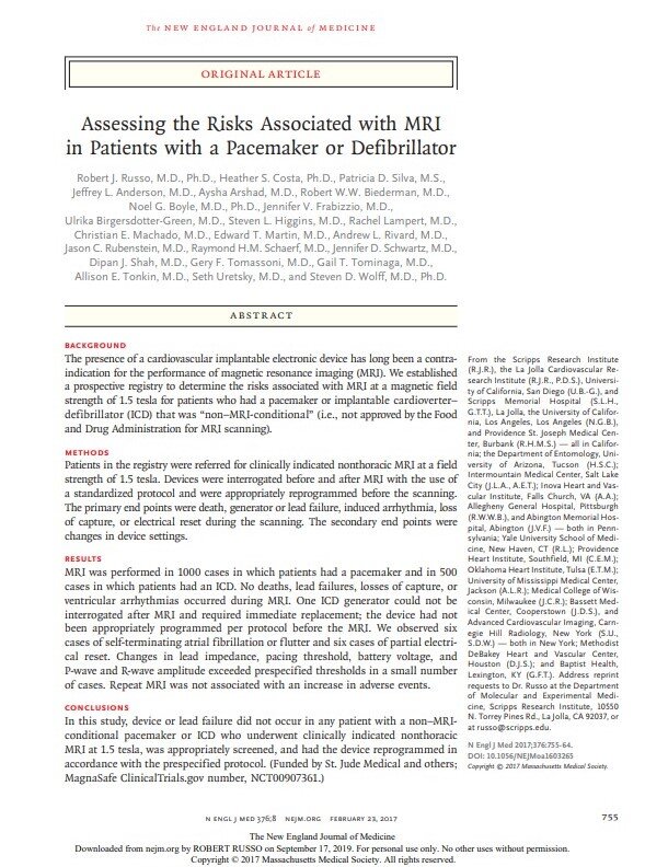
MagnaSafe Registry
Background: The presence of a permanent pacemaker or implanted cardioverter-defibrillator (ICD) has long been a contraindication to magnetic resonance imaging (MRI). However, the risk to both patient and device has never been sufficiently evaluated in a large-scale study.
Methods: Patients were referred for clinically-indicated non-thoracic MRI. Pacemaker and ICD interrogation was performed pre- and post-MRI using a standardized protocol. Devices were appropriately reprogrammed during the procedure. Pacemaker dependent ICD patients were excluded. Primary endpoints were death, generator/lead failure, induced ventricular or atrial arrhythmia or electrical reset. Secondary endpoints were device parameter changes.
Results: Clinically-indicated non-thoracic MRI was performed in 1500 patients (1000 pacemakers, 500 ICDs) at 21 clinical centers. No deaths, generator/lead failures, losses of capture, or ventricular arrhythmias occurred during the scan. One ICD generator required replacement when tachytherapy was inappropriately active during the exam. Six episodes of self-terminating atrial fibrillation (<49 hours) and 6 cases of partial electrical reset were observed. A change in pacing lead impedance of ≥50Ω occurred in 3% of pacemakers and 4% of ICDs. A decrease of ≥ 50% in P-wave amplitude occurred in 5 pacemakers and 1 ICD. A decrease of ≥25% in R-wave amplitude occurred in 4% of pacemakers and 2% of ICDs, and a decrease of ≥50% in 1 ICD. A pacing threshold increase ≥0.5V occurred in 1% of pacemaker and ICD leads.
Conclusions: Clinically-indicated non-thoracic MRI at 1.5T may be performed for patients with non-MRI conditional devices at no detectable risk when the device is appropriately programmed during the exam.
Eradicate Study
The purpose of the ERADICATE Study is to determine the radiation exposure for patients undergoing nuclear and non-nuclear cardiovascular stress-testing at a single institution between January 2003 and December 2012.
To determine the relative contribution of nuclear stress testing and diagnostic coronary angiography to the total diagnostic radiation exposure of a cardiovascular patient population over a 10 year period.
For patients undergoing cardiac stress testing the radiation exposure received from a nuclear-based stress test represents a significant portion (37%) of the cumulative diagnostic radiation exposure during a ten-year period (including cardiac and non-cardiac diagnostic procedures).
Conclusions: Adoption of a stress-first or stress-only protocol for nuclear-based cardiac stress testing results in a significant reduction in unnecessary radiation exposure for patients undergoing cardiac stress testing (from 37% to 25% of cumulative dose and is dependent on institutional SPECT normal rate).
MagnaSafe Pilot Study
Background: Despite recent research much controversy persists regarding the performance of MRI with a pacemaker or implantable cardioverter-defibrillator (ICD). MagnaSafe is a prospective registry designed to determine the risks of clinically-indicated MRI at 1.5-Tesla for patients with an implanted cardiac device.
Methods: Patients were referred for clinically-indicated non-thoracic MRI at 1.5 Tesla. Devices were interrogated pre- and post-MRI using a standardized protocol, and were appropriately reprogrammed during the scan. Primary endpoints were death, generator/lead failure, induced ventricular/atrial arrhythmia or electrical reset during the scan. Secondary endpoints were device parameter changes.
Results: MRI was performed in 1000 pacemakers and 500 ICDs at 21 centers. No deaths, lead failures, losses of capture, or ventricular arrhythmias occurred during the scan. One ICD generator required replacement when anti-tachytherapy was inappropriately active during MRI and the device became unresponsive. Six episodes of self-terminating atrial fibrillation and 6 cases of partial electrical reset were observed. For all devices a decrease of ≥ 50% in P-wave amplitude occurred in XX% of cases; a decrease of ≥25% in R-wave amplitude in YY%, and a decrease of ≥50% in 1; a pacing threshold increase ≥0.5V in ZZ% of leads; and a change in pacing lead impedance of ≥50Ω in AA%; which did not correlate with other parameter changes. Repeat MRI was not associated with an increase in adverse events.
Conclusions: Clinically-indicated non-thoracic MRI at 1.5 Tesla may be performed for patients with non-MRI conditional devices at no detectable risk when patients are appropriately screened and the device reprogrammed during the examination.
AVID
Background: Intravascular ultrasound (IVUS) is an invasive imaging technique used to visualize coronary cross-sectional anatomy. Several studies have examined the role of IVUS-guided stent placement with varying clinical restenosis rates. However, different IVUS criteria for optimal stent placement were used in each study. To test the hypothesis that IVUS-guided stent placement results in a lower rate of target lesion revascularization the present trial was initiated using prespecified IVUS criteria for procedure success.
Methods and Results: The Angiography Versus Intravascular ultrasound-Directed (AVID) stent placement study is a multicenter randomized clinical trial designed to determine the role of IVUS during elective coronary stent placement in native coronary arteries as well as bypass grafts. IVUS criteria for optimal stent placement (expansion, apposition and lack of dissection) had been previously tested in a pilot trial. Assessment of the primary endpoint was determined by clinical follow-up without repeat angiography. In an intention-to-treat analysis, the 12-month target lesion revascularization rate, which was the study’s primary outcome variable, was lower in the IVUS-directed treatment group compared to the angiography directed group (8.1% vs. 12.0%, P=0.08). In a per-protocol analysis, excluding protocol violations of a distal reference vessel of <2.5 mm, the 12-month rate of target lesion revascularization was 4.9% in the IVUS-directed group and 12.4% in the Angiography-guided group (P=0.01).
Conclusions: IVUS-directed stent placement results in larger acute stent dimensions without an increase in complications, and significantly lower 12-month TLR rates for vessels >2.5 mm by angiography, vein grafts, and vessels with high-grade preprocedure stenosis.
The Left Main IVUS Registry
The Left Main IVUS Registry Results: A significant left main stenosis was found in 47% of patients with an inconclusive angiogram who were then eligible for CABG/Left Main PCI.
Purpose: To determine if IVUS is superior to angiography for the evaluation of inconclusive angiogram in the setting of minimal disease by angiography (<50% stenosis).
For patients with an inconclusive coronary angiogram (<50%), a significant stenosis was identified in 33% of patients by IVUS.
Angiographic lesion location, diameter, and calcium content were predictive of a significant stenosis when an inconclusive lesion was evaluated by IVUS.
Lesion severity by angiography was not predictive of a significant stenosis detected by IVUS (and need for revascularization).
When angiography is non-diagnostic (stenosis <50%), IVUS is an essential technique for the identification of clinically important left main coronary disease.
STAT Registry
A multicenter registry (Stent Thrombosis After Ticlopidine therapy) of bare-metal stent thrombosis (acute and delayed) for patients treated with Ticlopidine.
Porcine Model of Restenosis
Background: To determine if an aggressive approach to coronary revascularization with oversized balloons is counterproductive, we studied the effect of balloon-to-artery (B:A) ratios on neointimal hyperplasia following stent placement in porcine coronary arteries.
Methods and Results: A porcine coronary overstretch model was used to determine the effect of varying B:A ratios on neointimal growth after intravascular ultrasound (IVUS)-guided primary stent placement. Vessels were randomly assigned to one of five B:A ratios between 1.0 and 1.4 to 1. IVUS imaging was performed pre- and post-stent placement and at 28-day follow-up. Quantitative IVUS measurements of stent, vessel lumen, and neointimal diameter and area were made every 1 mm along the stent length. Volumes were calculated using Simpson’s rule. Sixty vessels in 33 animals were treated with coronary stent placement. Stent recoil increased with increasing B:A ratio (linear regression test for slope, P=.003). Neointimal volume increased with increasing B:A ratio (P<0.001) and was independent of vessel size. Even minor vessel overstretch (B:A ratio of 1.1-to-1) resulted in neointimal hyperplasia. In-stent volume stenosis and maximum cross-sectional area stenosis also increased with increasing B:A ratio (P=.001 and P<.001, respectively) and were independent of vessel . Neointimal growth extended beyond the stent edge (2.58±2.09 mm), increased with increasing B:A ratio (P<.001), and was independent of vessel size.
Conclusion: In a porcine model of IVUS-guided primary stent placement, in-stent neointimal volume was strongly associated with increasing balloon-to-artery ratio and was independent of vessel size. The clinical implication is that careful matching of vessel and stent diameters by IVUS may minimize neointimal hyperplasia.


